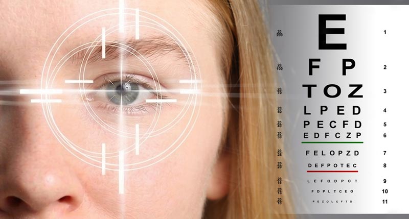
If you’ve had an eye exam recently — and we hope you have! — You know that eye exams are not what they used to be. If you think an eye exam is all about reading the letters on the Snellen chart, you’re in for an … ahem … eye-opening experience in our offices.
Modern eye exams are faster, more comfortable, and far more powerful than most patients realize. Some of the most serious eye diseases begin without warning. But with today’s cutting-edge technological tools that make eye exams more accurate, your optometrist can detect early signs of disease, monitor changes over time, and help protect your vision with greater precision. Plus, these game-changing modern tools and tests make eye exams more thorough and comfortable.
Let’s take a look at some new technologies that your optometrist may use to diagnose, treat, and preserve your eyesight.
- Optical Coherence Tomography (OCT) Scans
Think of this technology like having an “MRI” of your eye. OCT scans use non-invasive light waves to provide cross-sectional, three-dimensional images of the retina and optic nerve. This provides a detailed analysis of the tiny structures in each layer of ocular tissue. It can aid your optometrist in detecting subtle nerve fiber loss seen in many types of glaucoma and also can identify early signs of diabetic retinopathy or macular degeneration. OCT technology goes beyond what your optometrist can see during a traditional eye exam to provide a more precise diagnosis and personalized treatment plan.
- Fundus Photography
The fundus is the inside surface at the back of your eye, and it is a critical part of your vision. Fundus photography can be a part of your routine eye exam. It is a simple, non-invasive process using a fundus camera to capture a high-resolution, colored, or specialized image of the retina to diagnose and treat ocular disease. Your optometrist may give you eye drops to dilate your pupils before taking the photos. The camera captures images of the retina, optic nerve, macula, choroid, vitreous, and retinal blood vessels to detect preventable and treatable eye diseases and serves as a baseline record for monitoring disease progression.
Fundus photography is extremely safe and has no side effects other than the after-effects of dilated pupils, which may make your eyes sensitive to light for a few hours.
- Visual Field Testing
Your peripheral vision is just as important as central vision! This test measures how far you can see on the sides and outer edges of your vision, and can also identify any blind spots you may have. Visual Field Testing is a crucial tool for diagnosis because most types of glaucoma first affect your peripheral vision. Visual field testing allows your optometrist to track changes to your vision over time and assess how well treatments are working.
- Corneal Pachymetry
This test in a non-invasive ultrasonic technique to measure corneal thickness, which is useful in evaluating intraocular pressure. It measures the transparent, tough tissue that covers the pupil and iris. This tissue helps you focus and offers your eyes protection from dirt and bacteria.
Corneal Pachymetry is used primarily for patients at risk for glaucoma, refractive errors, or corneal disease. Having a thicker cornea may mean that you are less likely to develop glaucoma and is also an indicator of eye pressure. Knowing the thickness of the corneal tissue can help your optometrist diagnose you more precisely.
- Corneal Topography
This test is valuable for patients who wear contact lenses or those considering LASIK. It’s an essential part of pre-operative planning for LASIK and other surgeries because it creates a three-dimensional, detailed map of the cornea’s surface, identifying shape, curvature, and irregularities that affect your vision quality. Corneal Topography essentially charts the landscape of your cornea – both its steepness (called keratoconus) or flatness — to assess corneal astigmatism, refractive power, and other abnormalities.
It’s a quick and painless test; nothing touches your eye. It produces a series of color-coded maps that your optometrist uses to diagnose and manage various eye conditions. Patients who wear contact lenses benefit from the data gathered because it ensures enhanced comfort, vision clarity, and a better fit.
- Tear Film Quality Assessment
Your optometrist can assess tear film quality through a variety of methods, ranging from simple staining and break-up time to more elaborate screening devices. Tear film is the thin lipid layer covering the surface of the eye that provides protection, lubrication, and nourishment for the eye.
Many eye doctors use the Schirmer Strip, a test requiring no anesthesia, to diagnose conditions such as Dry Eye Syndrome. The Schirmer Strip evaluates the amount of tears absorbed by a thin strip of filter paper. For the test, an absorbent test strip is draped over the lower lid, then after 5 minutes, the wetting length is measured, helping your eye doctor determine whether you are experiencing Dry Eye Syndrome.
These state-of-the-art technologies and tools transform the way we safeguard your vision. Your optometrist can now examine your eyes with more precision and comfort, delivering the proactive care that you deserve.
Contact our office and schedule your next eye exam today to let modern technology provide added benefits to your long-term vision health.